
PROMO!
First order? Get 10% OFF with this code: 1storder


Written by

Research shows the IGF-1 pathway plays an important role in muscle recovery, protein synthesis, and muscle growth. IGF-1 LR3 is a modified form of insulin-like growth factor-1 that stays active longer than natural IGF-1.
It also binds less strongly to IGF-binding proteins. Because of these properties, it is widely studied in muscle recovery research. Studies suggest IGF-1 LR3 may increase protein synthesis, activate satellite cells, and support muscle regeneration through IGF-1 receptor signaling. Most of this evidence comes from laboratory and animal studies.
This guide explains what current research says about IGF-1 LR3 as a muscle recovery peptide. It explores how IGF-1 LR3 supports muscle repair and protein synthesis, and how it compares with other recovery peptides.
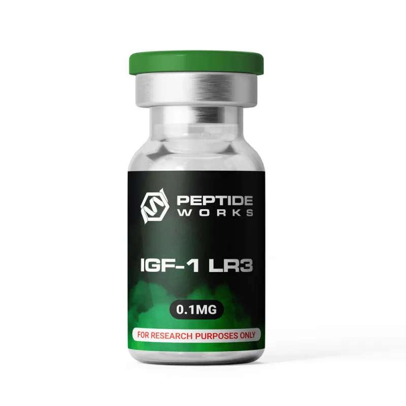
Explore IGF-1 LR3 from Peptide Works, a peptide that boosts muscle protein synthesis and accelerates quicker recovery for enhanced strength.
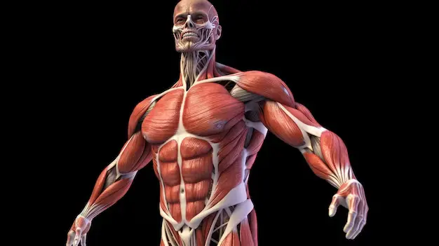
IGF-1 LR3 supports protein synthesis by activating the IGF-1 receptor and the PI3K/Akt/mTOR signaling pathway in muscle cells. Research shows this pathway increases protein synthesis and promotes muscle cell growth. IGF-1 LR3 also has reduced binding to IGF-binding proteins, which allows it to remain active longer than natural IGF-1.
Studies also suggest IGF-1 signaling activates satellite cells that help repair and regenerate damaged muscle fibers. Unlike BPC-157 and TB500, which are studied mainly for tissue repair through different biological pathways, IGF-1 LR3 is primarily studied for its role in muscle growth and anabolic signaling. Its longer activity profile has made it a common peptide for research on muscle recovery and regeneration.
Protein synthesis is regulated by the mTOR pathway. which controls muscle growth and repair in response to nutrients and growth signals. Because IGF-1 LR3 activates this pathway, researchers continue to study its potential role in supporting muscle regeneration and recovery after muscle injury.
Discover BPC-157 from Peptide Works, a peptide that promotes tendon, ligament, and tissue repair for faster injury recovery.
mTOR acts as the master control switch for muscle building and recovery processes. This cellular pathway decides when muscles grow or break down based on available nutrients and signals. Recovery peptides like IGF-1 LR3 directly activate mTOR to boost protein creation. The pathway turns on ribosome production and amino acid uptake for muscle repair. mTOR also controls autophagy, which cleans damaged proteins from muscle cells.
BPC-157 and TB500 work through different pathways but still influence mTOR activity indirectly. When mTOR gets activated, muscles switch from breakdown mode to building mode. This makes mTOR the key target for effective recovery peptide action.
Understanding how mTOR controls these processes leads to an important question about timing how quickly do these effects actually occur?
The speed of recovery peptide activity depends on the biological pathway being studied rather than a fixed timeline. Animal studies show that IGF-1 signaling can rapidly activate protein synthesis and reduce protein breakdown after muscle injury.
Studies also report increased satellite cell activity and faster muscle regeneration, but they do not establish how many days or weeks are required for measurable muscle growth.
BPC-157 and TB500 work through different repair pathways. Research suggests BPC-157 may support tissue repair while TB500 is studied for its role in cell migration and tissue regeneration.
Current studies do not show how quickly these peptides affect muscle growth. More research is needed to understand how they support muscle recovery over time.
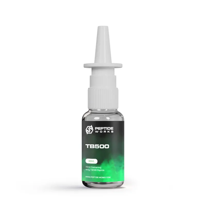
Checkout TB500 from Peptide Works, a peptide that supports cell migration and reduces inflammation for comprehensive tissue healing.
Recovery peptides support tissue repair through different biological pathways. Animal studies show that BPC-157 promotes angiogenesis, increases collagen synthesis, and stimulates fibroblast activity. These effects help repair tendons, ligaments, skeletal muscle, and other connective tissues.
Studies also show that BPC-157 regulates nitric oxide signaling and reduces inflammatory cytokines during the healing process. TB500 (Thymosin β4) supports tissue repair by promoting cell migration, tissue remodeling and inflammation resolution.
IGF-1 LR3 supports muscle repair by activating the IGF-1 receptor and the PI3K/Akt/mTOR pathway, which increases protein synthesis and activates satellite cells involved in muscle regeneration. Because these peptides work through different mechanisms, researchers continue to study their potential roles in muscle and soft tissue repair.
While these recovery benefits are well-documented, researchers should also understand the potential side effects before implementing these compounds.
Animal studies suggest most recovery peptides are well tolerated. However, side effects can vary by peptide, and long-term safety is still being studied.
Potential side effects reported in preclinical studies include:
Current animal studies report few serious side effects, but more research is needed to better understand the long-term safety of these recovery peptides.
IGF-1 LR3, BPC-157, and TB500 are studied for different areas of tissue repair and muscle recovery. Current research does not show that one peptide works better than the others. Their effects depend on the biological pathway being studied and the type of tissue involved.
| Recovery Peptide | Primary Research Focus | Main Mechanism |
| IGF-1 LR3 | Muscle regeneration | IGF-1 receptor signaling and anabolic pathways |
| BPC-157 | Connective tissue repair | Collagen formation and angiogenesis |
| TB500 (Thymosin β4) | Tissue remodeling | Cell migration and inflammation regulation |
As research in this field continues to expand, the future holds even greater potential for recovery peptide applications.
IGF-1 LR3 is a strong muscle recovery peptide that boosts protein building through cellular pathways. Comparing IGF-1 LR3, BPC-157, and TB500 shows how each peptide works differently for recovery needs. These compounds work in different ways – from building muscle to fixing tissue and reducing swelling.
Current studies show good results with mild side effects when used right. The future of muscle recovery peptides looks bright as scientists keep making better compounds with safer profiles.
Better delivery methods and mixing peptides may soon give faster recovery times and better results for researchers studying muscle repair and tissue healing. Peptide Works provides research-grade compounds for scientific investigation into these promising recovery applications.
All products discussed are supplied for research purposes only and are not intended for human use.
(1) Philippou A, Barton ER. Optimizing IGF-I for skeletal muscle therapeutics. Growth Horm IGF Res. 2014 Oct;24(5):157-63.
(2) Song YH, Song JL, Delafontaine P, Godard MP. The therapeutic potential of IGF-I in skeletal muscle repair. Trends Endocrinol Metab. 2013 Jun;24(6):310-9.
(3) Pevec D, Novinscak T, Brcic L, Sipos K, et al. Impact of pentadecapeptide BPC 157 on muscle healing impaired by systemic corticosteroid application. Med Sci Monit. 2010 Mar;16(3):BR81-88.
(4) Vasireddi N, Hahamyan H, Salata MJ, Karns M, et al. Emerging Use of BPC-157 in Orthopaedic Sports Medicine: A Systematic Review. HSS J. 2025 Jul 31:15563316251355551.
(5) Spurney CF, Cha HJ, Sali A, Pandey GS, et al. Evaluation of skeletal and cardiac muscle function after chronic administration of thymosin beta-4 in the dystrophin deficient mouse. PLoS One. 2010 Jan 29;5(1):e8976.
ALL CONTENT AND PRODUCT INFORMATION AVAILABLE ON THIS WEBSITE IS FOR EDUCATIONAL PURPOSES ONLY.
DISCLAIMER: These products are intended solely as a research chemical only. This classification allows for their use only for research development and laboratory studies. The information available on our Peptide Works website: https://peptide-works.com/ is provided for educational purposes only. These products are not for human or animal use or consumption in any manner. Handling of these products should be limited to suitably qualified professionals. They are not to be classified as a drug, food, cosmetic, or medicinal product and must not be mislabelled or used as such.
Peptide Works
Related Articles
How can ARA-290 Peptide Help Diabetic Patients?
Diabetes affects millions of people around the world and leads to serious health problems. Research shows that the ARA-290 peptide

Can AICAR Peptide Improve Endurance?
AICAR peptide has shown promising results for improving endurance capacity in studies. Research shows that AICAR activates AMPK, increasing glucose

Enhancing Energy with NAD+ Supplements
Energy is the driving force behind focus, movement, and recovery. When cells have less energy to work with, the body